Imaging Evaluation of Nephrolithiasis
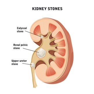
CT KUB (Non-Contrast CT) Secondary Signs of obstruction X-ray KUB Intravenous Pyelogram (IVP) Final Take-Home Points Key Imaging Findings: Renal or ureteric calculus appears as a hyperdense focus (usually >200 HU). Detects stones as small as 1–2 mm. Precisely defines size, number, and exact location. Common sites of impaction: PUJ, iliac crossing, VUJ. Stone Characterization […]