
PATIENT SNAPSHOT
40 YEAR-OLD MALE
CHIEF COMPLAINTS
- Headache
- Blurring of vision
HISTORY OF PRESENTING ILLNESS
- Headache — insidious onset, gradually progressive over several months; dull-aching and holocranial in character, more pronounced in the early morning hours; partial relief with over-the-counter analgesics.
- Visual disturbance — gradual, bilateral blurring of vision with peripheral field involvement noticed by the patient (“bumping into people or objects on either side”), progressively worsening and prompting the current hospital visit.
- No history of head trauma, seizures, vomiting, or loss of consciousness at presentation.
MASS-EFFECT SCREEN
- Diplopia — none reported
- Vomiting — absent, no projectile vomiting
- Seizures or focal weakness — none
- No early-morning worsening beyond the headache itself
ENDOCRINE SCREEN
- Galactorrhea / gynecomastia — absent
- Libido / erectile function — no change reported
- Polyuria / polydipsia — absent
- Acral or facial coarsening (acromegaly) — none noted
- Weight gain, easy bruising, proximal weakness (Cushing’s) — none noted
Note:
No significant past medical or surgical history. No family history of pituitary or other endocrine disease.
Examination | Finding |
General survey | Conscious, oriented; vitals stable; no pallor or icterus |
Visual acuity | Mildly reduced, bilateral |
Visual fields (confrontation / perimetry) | Bitemporal hemianopia — pattern of optic chiasm compression |
Fundus examination | Bilateral optic disc pallor |
Cranial nerves III, IV, VI | Grossly intact; no ptosis or diplopia |
Motor, sensory, cerebellar exam | Unremarkable |
General stigmata | No features of acromegaly, Cushing’s syndrome, or hypothyroidism |
Differential Diagnosis
Given the patient’s age and the bitemporal-hemianopia pattern, sellar / parasellar masses were the leading clinical differentials, ranked as follows:
- Pituitary macroadenoma — most likely, given the patient’s age and the chiasmal compression pattern on examination.
- Craniopharyngioma
- Suprasellar / tuberculum sellae meningioma
- Rathke’s cleft cyst
- Optic or hypothalamic glioma — less likely at this age.
- Sellar metastasis — less likely, no known primary malignancy.
LABORATORY — HORMONAL PROFILE
- CBC, renal & liver function (baseline)
- Serum prolactin
- Growth hormone / IGF-1
- 8 AM serum cortisol & ACTH
- TSH, free T4
- FSH, LH, testosterone
IMAGING & FUNCTIONAL TESTS
- Visual field perimetry
- Contrast-enhanced MRI brain — dedicated pituitary protocol
- (Investigation of choice for sellar/parasellar masses)
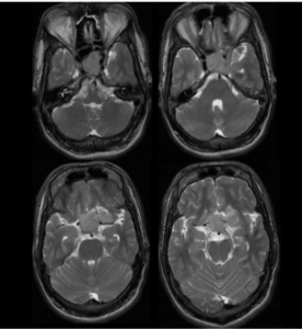
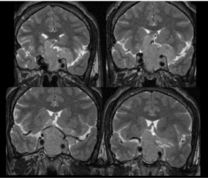
ORONAL T2
AXIAL T2
- A well-defined, heterogeneously hyperintense mass is centered in the sellar region.
- The mass extends superiorly into the suprasellar cistern, abutting and elevating the optic chiasm.
- Maximum diameter exceeds 1 cm — in keeping with a macroadenoma rather than a microadenoma.

Sellar–suprasellar extension
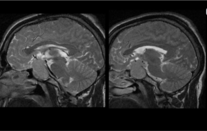
Sagittal sections confirm that the mass extends from the sella into the suprasellar cistern
MRI Brain — Sagittal View
- Chiasmal involvement
The lesion abuts the floor of the third ventricle and elevates the optic chiasm — explaining the patient’s visual symptoms
- Posterior extent
No obvious extension into the posterior fossa or brainstem
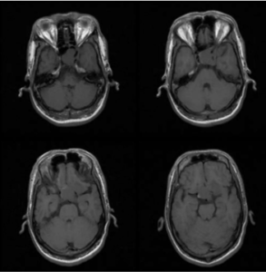
- On pre-contrast T1-weighted images, the sellar mass blends with adjacent brain parenchyma and is poorly conspicuous
- Following contrast administration, the lesion shows distinct heterogeneous enhancement that clearly outlines its margins — confirming a vascularised sellar mass
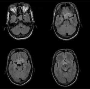
- Chiasm relationship
Best demonstrates the relationship of the mass to the optic chiasm above and the cavernous sinuses on either side
- Gland of origin
The normal pituitary gland is not separately identified, in keeping with an adenoma arising from and replacing the gland
- Note
For complete vascular and cavernous-sinus characterization, correlate with the formal radiology report
Sellar–suprasellar mass lesion, heterogeneous on T2-weighted images with heterogeneous post-contrast enhancement, causing superior displacement and compression of the optic chiasm — findings consistent with a Pituitary Macroadenoma.
Functional status (secretory vs non-secretory) to be determined by hormonal profile
Sellar–suprasellar mass, > 10 mm, with optic chiasm compression syndrome
PENDING HORMONAL CORRELATION
What Is a Pituitary Macroadenoma?
Definition
- A benign, monoclonal tumor of the anterior pituitary gland (adenohypophysis).
- Among the most common intracranial neoplasms — roughly 10–15% of all primary brain tumors.
- The overwhelming majority occur sporadically; a small subset is associated with MEN type 1 syndrome.
CLASSIFICATION BY SECRETORY ACTIVITY
FUNCTIONING (HORMONE-SECRETING)
- Prolactinoma (most common)
- GH-secreting (acromegaly)
- ACTH-secreting (Cushing’s disease)
- TSH-secreting (rare)
NON-FUNCTIONING
- No hormone hypersecretion; the tumor presents because of mass effect alone (headache, visual field loss, hypopituitarism).
Mass Effect?
- Headache
- Bitemporal visual field loss / blurring (chiasm compression)
- Cranial nerve III, IV, VI palsy — if cavernous sinus is invaded
- Hypopituitarism — fatigue, hypogonadism, hypothyroidism
Harmonal Hypersectretion
- Galactorrhea, amenorrhea — prolactinoma
- Acral / facial coarsening, increased shoe or glove size — acromegaly
- Central obesity, purple striae, easy bruising — Cushing’s disease
1. Evaluate
- Multidisciplinary work-up — endocrinology, neurosurgery, ophthalmology
2. Classify
- Complete hormonal profile to identify functioning vs non-functioning tumor
3. Prolactinoma
- First-line medical therapy with dopamine agonists (cabergoline / bromocriptine); surgery reserved for resistance or intolerance
4. Non-functioning + Mass Effect
- Trans-sphenoidal endoscopic surgery (first choice); craniotomy if extensive lateral/suprasellar extension
5. GH- / ACTH-secreting
- Surgery first-line; somatostatin analogues or other medical therapy as adjunct
6. Post-operative Care
- Hormone replacement for hypopituitarism; monitor for diabetes insipidus
7. Residual / Recurrent Disease
- Stereotactic radiosurgery or fractionated radiotherapy
- Visual recovery is generally favorable with timely chiasmal decompression, before chronic optic atrophy sets in
- Serial MRI surveillance (e.g., 3 months post-operatively, then annually) to detect residual or recurrent tumor
- Periodic hormonal assessment; lifelong hormone replacement may be required in some patients
- Overall prognosis is favorable for non-functioning macroadenomas with timely surgical decompression