Each week, an interesting radiology case is selected and shared with members. The case includes anonymized patient history, imaging findings, and key clinical questions. Members are encouraged to analyze the images, suggest possible diagnoses, and discuss the best imaging approach. A faculty moderator or senior resident guides the discussion, providing expert insights into differential diagnoses, imaging techniques, and case management. This activity ensures continuous learning and improves radiological pattern recognition skills, ultimately fostering a culture of analytical thinking among radiology residents.
Radiology is a cornerstone of modern medicine, offering critical insights that guide diagnosis and treatment across all specialties. The Radiology Case of the Week is an engaging initiative designed to enhance diagnostic skills through real-world imaging cases. Each week, we present an interesting case covering various imaging modalities, including X-ray, CT, MRI, ultrasound, and nuclear medicine, along with key findings, differential diagnoses, and clinical correlations.
Whether you are a medical student, resident, or radiology enthusiast, this is an opportunity to sharpen your interpretation skills, challenge your diagnostic reasoning, and stay updated with radiology best practices. Join us in exploring complex cases, discussing imaging patterns, and learning from expert insights.
Stay tuned, participate, and elevate your radiology expertise—one case at a time!
Dates to be communicated to members as per schedule
Introduction
In the rapidly evolving field of radiology, proficiency in interpreting CT (Computed Tomography) and MRI (Magnetic Resonance Imaging) images is paramount for healthcare professionals. The ability to accurately analyze these imaging modalities enables clinicians to diagnose conditions effectively and formulate appropriate treatment plans. This workshop, organized by the RADNOVA radiology club, aims to enhance participants’ skills in interpreting CT and MRI images using DICOM (Digital Imaging and Communications in Medicine) viewers.
This comprehensive session will delve into the intricacies of image interpretation, focusing on the principles underlying CT and MRI technologies, the practical aspects of utilizing DICOM viewers, and the critical competencies required for effective image analysis.
Workshop Objectives
The primary objectives of this workshop are as follows:
- Understanding Imaging Modalities: Participants will gain a solid foundation in the principles of CT and MRI imaging, including their respective advantages, limitations, and clinical applications.
- Familiarization with DICOM Standards: Attendees will be introduced to the DICOM standard, which facilitates the sharing and management of medical imaging data, ensuring interoperability among different imaging devices and software.
- Hands-On Experience with DICOM Viewers: Participants will engage in hands-on sessions using DICOM viewers to explore various imaging studies, honing their skills in image manipulation and interpretation.
- Developing Analytical Skills: Through guided exercises and case discussions, attendees will learn to recognize common pathological findings, differentiate between normal and abnormal images, and enhance their overall interpretive skills.
- Integration of Clinical Context: The workshop will emphasize the importance of integrating clinical information with imaging findings to arrive at accurate diagnoses.
Workshop Outline
- Understanding Imaging Modalities
CT and MRI are essential imaging techniques that play a crucial role in modern diagnostic medicine.
CT Imaging:
Principles: CT imaging utilizes X-rays to generate cross-sectional images of the body. The X-ray tube rotates around the patient, capturing multiple images from different angles, which are then reconstructed to produce detailed images of internal structures.
Advantages: CT is particularly effective in visualizing bony structures, detecting acute hemorrhages, and assessing complex anatomical regions such as the abdomen and pelvis.
Limitations: The use of ionizing radiation poses a risk, necessitating a careful evaluation of the risks versus benefits. Additionally, CT is less effective in differentiating soft tissue contrast compared to MRI.
MRI Imaging:
Principles: MRI employs strong magnetic fields and radiofrequency pulses to generate images based on the magnetic properties of hydrogen atoms in the body. This technique produces high-resolution images of soft tissues.
Advantages: MRI excels in visualizing soft tissue structures, making it invaluable for assessing neurological, musculoskeletal, and oncological conditions.
Limitations: MRI is time-consuming, more expensive than CT, and may not be suitable for patients with certain implants or devices.
- Familiarization with DICOM Standards
The DICOM standard is essential for managing and sharing medical imaging data. Participants will learn:
DICOM Structure: Understanding the DICOM file structure, which includes metadata (patient information, imaging modality, acquisition parameters) and image pixel data.
Interoperability: The importance of DICOM in ensuring compatibility among various imaging devices and software systems, facilitating seamless data exchange.
DICOM Viewers: An overview of various DICOM viewers available for image analysis, including open-source options, commercial software, and web-based platforms.
- Hands-On Experience with DICOM Viewers
The hands-on segment of the workshop will involve interactive sessions where participants will use DICOM viewers to analyze a range of CT and MRI studies.
Image Manipulation Techniques:
Windowing and Leveling: Adjusting brightness and contrast to optimize the visualization of specific tissues.
Image Reconstruction: Understanding the reconstruction process, including 3D reconstructions and MPR (multiplanar reconstructions).
Annotation Tools: Utilizing tools for measuring distances, angles, and regions of interest (ROIs) on images.
Case Studies: Participants will be provided with a selection of clinical cases representing common and challenging scenarios in CT and MRI interpretation. They will work in groups to analyze the images, identify key findings, and discuss differential diagnoses.
- Developing Analytical Skills
Effective image interpretation requires a systematic approach. The workshop will cover:
Recognizing Normal Anatomy: Establishing a baseline understanding of normal anatomical structures as a foundation for identifying abnormalities.
Identifying Common Pathologies: Training participants to recognize prevalent conditions, such as tumors, fractures, and degenerative changes, by analyzing case examples.
Critical Thinking: Encouraging participants to develop their analytical skills through guided discussions and peer interactions, fostering a collaborative learning environment.
- Integration of Clinical Context
The final segment of the workshop will focus on integrating imaging findings with clinical context:
Clinical Correlation: Emphasizing the importance of correlating imaging results with clinical symptoms, laboratory findings, and patient history to arrive at accurate diagnoses.
Discussion of Real-World Cases: Engaging participants in discussions of real-world clinical cases where imaging played a pivotal role in diagnosis and management, highlighting the necessity of collaboration between radiologists and referring physicians.
- Conclusion
The CT & MRI Image Interpretation workshop, hosted by the RADNOVA radiology club, is designed to equip participants with essential skills and knowledge in image interpretation. By combining theoretical knowledge with practical experience using DICOM viewers, attendees will leave the workshop with enhanced confidence in their ability to analyze CT and MRI images effectively. This workshop not only aims to improve technical competencies but also fosters a deeper understanding of the vital role radiology plays in patient care.
Through this session, we hope to inspire the next generation of radiologists and healthcare professionals to continue pursuing excellence in the field of medical imaging. As the landscape of radiology evolves, ongoing education and hands-on experience will remain critical in ensuring that practitioners are well-prepared to meet the challenges of modern medicine.
- References
American College of Radiology (ACR). (2023). ACR Appropriateness Criteria.
DICOM Standards Committee. (2023). DICOM Standard.
Brant, W. E., & Helms, C. A. (2012). Fundamentals of Diagnostic Radiology (4th ed.). Lippincott Williams & Wilkins.
Latchaw, R. E., et al. (2009). “American College of Radiology White Paper on Imaging and Informatics.” Journal of the American College of Radiology
Dates to be communicated to members as per schedule
Introduction
Interventional Radiology (IR) is a subspecialty of radiology that utilizes minimally invasive procedures to diagnose and treat a variety of medical conditions. By employing imaging guidance techniques, such as fluoroscopy, ultrasound, CT, and MRI, interventional radiologists can perform a wide range of procedures with precision and reduced patient risk. This workshop, organized by the RADNOVA radiology club, aims to provide participants with a comprehensive introduction to basic interventional radiology procedures, such as biopsies and catheter placements.
The workshop will cover the fundamental principles of interventional radiology, explore various IR techniques and their clinical applications, and provide hands-on training in basic procedures. By the end of the session, participants will be equipped with essential knowledge and skills to appreciate the role of IR in modern medicine and enhance their competency in performing basic IR procedures.
Workshop Objectives
The primary objectives of this workshop are as follows:
- Understanding Interventional Radiology: Participants will gain an understanding of the principles and practices of interventional radiology, including the various imaging modalities used in IR.
- Familiarization with Basic Procedures: Attendees will learn about common interventional radiology procedures, such as biopsies, catheter placements, and drainage procedures, including their indications and contraindications.
- Hands-On Experience: Participants will engage in hands-on training sessions to practice basic IR techniques using simulators or models, enhancing their procedural skills.
- Exploration of Clinical Applications: The workshop will emphasize the clinical applications of IR procedures in various medical fields, including oncology, vascular disease, and gastroenterology.
- Patient Safety and Ethical Considerations: Participants will learn about the importance of patient safety, informed consent, and ethical considerations in interventional radiology practice.
Workshop Outline
- Understanding Interventional Radiology
Interventional radiology has transformed the landscape of modern medicine by providing minimally invasive alternatives to traditional surgical procedures. Participants will be introduced to the following topics:
Definition and History of Interventional Radiology:
Overview of the evolution of interventional radiology as a subspecialty, highlighting key milestones in its development and the introduction of various imaging technologies.
Principles of Image Guidance:
Understanding the role of imaging modalities in guiding interventional procedures, including:
Fluoroscopy: Real-time X-ray imaging for visualizing dynamic processes, particularly in vascular interventions.
Ultrasound: Utilizing sound waves to visualize soft tissue structures and guide needle placement in biopsies and drainage procedures.
CT and MRI: Providing high-resolution imaging for accurate localization of lesions and guiding complex interventions.
The Role of Interventional Radiologists:
Exploring the multifaceted roles of interventional radiologists in healthcare, including diagnosis, treatment, and collaboration with other specialties.
- Familiarization with Basic Procedures
This section will focus on the most commonly performed interventional radiology procedures, their indications, contraindications, and techniques. Participants will learn about:
Biopsy Procedures:
Indications: Understanding when biopsies are necessary for diagnosing malignancies or infections.
Techniques: Overview of various biopsy techniques, including fine-needle aspiration (FNA), core needle biopsy, and image-guided biopsies.
Procedural Steps:
Patient preparation, including obtaining informed consent and ensuring appropriate imaging guidance.
Techniques for local anesthesia and sedation, as well as the use of ultrasound or CT for needle localization.
Catheter Placement Procedures:
Types of Catheters: Understanding various catheter types, including central venous catheters (CVCs), peripherally inserted central catheters (PICCs), and drainage catheters.
Indications: Situations where catheter placement is indicated, such as for chemotherapy, long-term medication administration, or drainage of abscesses.
Procedural Steps:
Selection of appropriate access site and imaging guidance for catheter placement.
Techniques for catheter insertion, securing the catheter, and post-procedure care.
Drainage Procedures:
Indications: Understanding when percutaneous drainage is necessary for abscesses, fluid collections, or biliary obstruction.
Techniques: Overview of drainage techniques and the importance of imaging guidance to ensure accurate placement.
- Hands-On Experience
The hands-on portion of the workshop is designed to provide participants with practical experience in basic interventional radiology procedures. Participants will engage in simulated training sessions where they will:
Practice Biopsy Techniques:
Utilizing models or simulators to practice performing biopsies, including needle insertion, localization, and obtaining tissue samples.
Learn Catheter Placement Skills:
Engaging in hands-on training for catheter placement using simulation models, focusing on proper technique, securing the catheter, and troubleshooting common issues.
Explore Drainage Procedures:
Simulating drainage procedures to gain proficiency in identifying fluid collections and placing drainage catheters.
Feedback and Assessment:
Participants will receive feedback on their techniques from experienced instructors, enhancing their skills and confidence in performing these procedures.
- Exploration of Clinical Applications
This section of the workshop will focus on the various clinical applications of interventional radiology procedures in different medical fields:
Oncology:
The role of interventional radiology in cancer diagnosis and treatment, including tumor biopsies, radiofrequency ablation, and transarterial chemoembolization (TACE).
Vascular Disease:
Understanding how IR procedures are used to treat vascular conditions, such as peripheral artery disease, thrombolysis for deep vein thrombosis, and stenting.
Gastroenterology:
Exploring the use of interventional radiology in managing gastrointestinal conditions, including biliary drainage, portosystemic shunts, and feeding tube placements.
Urology:
The applications of IR in urological conditions, such as nephrostomy tube placement and renal biopsy.
Emergencies:
Discussing the critical role of interventional radiology in emergency settings, including trauma management and acute gastrointestinal bleeding interventions.
- Patient Safety and Ethical Considerations
Patient safety is paramount in interventional radiology. This segment will address:
Risk Assessment and Management:
Understanding potential complications associated with interventional procedures and strategies for minimizing risks.
Informed Consent:
The importance of obtaining informed consent, ensuring that patients understand the procedure, its risks, and benefits.
Ethical Considerations:
Discussing the ethical responsibilities of interventional radiologists, including patient autonomy, beneficence, and non-maleficence.
Quality Improvement and Best Practices:
Emphasizing the need for continuous quality improvement in interventional radiology, including adherence to guidelines and protocols to enhance patient safety.
Conclusion
The Basic Interventional Radiology workshop organized by the RADNOVA radiology club serves as an essential platform for medical students, residents, and healthcare professionals to gain a foundational understanding of interventional radiology procedures. By integrating theoretical knowledge with hands-on experience, participants will enhance their skills and confidence in performing basic IR techniques, such as biopsies and catheter placements.
This workshop highlights the significance of interventional radiology as a critical component of modern medicine, illustrating its diverse applications across various clinical specialties. As the field of interventional radiology continues to evolve, ongoing education and training will remain vital in ensuring practitioners are equipped to provide safe and effective care to their patients.
References
American College of Radiology (ACR). (2023). ACR Appropriateness Criteria.
Sacks, D., et al. (2010). “Interventional Radiology: A Guide to the Specialty.” American Journal of Roentgenology.
Dake, M. D., et al. (2017). “Interventional Radiology: Clinical Applications.” In Interventional Radiology: A Practical Guide (pp. 1-15). Springer.
Becker, G. J., & Kuhlman, J. E. (2016). “Safety and Effectiveness of Interventional Procedures.” Radiology.
Nascimento, F. A., et al. (2018). “The Role of Interventional Radiology in Emergency Medicine.” Emergency Medicine Clinics of North America.
Radiology is a constantly evolving field, with advancements in imaging technology, artificial intelligence, and interventional techniques shaping the future of medical diagnostics and treatment. Guest Lectures by Leading Radiologists provide a unique opportunity to learn from renowned experts in the field, gaining insights into cutting-edge developments, best practices, and career guidance.
Eminent radiologists from academic institutions and private practice are invited to share their expertise on subspecialties such as neuroradiology, musculoskeletal imaging, interventional radiology, and breast imaging. These lectures highlight advancements in imaging modalities, discuss complex cases, and provide career guidance to aspiring radiologists. Through direct interactions with field experts, members gain an understanding of emerging technologies, radiology best practices, and potential career paths within the specialty.
Each lecture features distinguished radiologists sharing their expertise on topics such as advanced imaging techniques, subspecialty innovations, AI integration in radiology, and real-world case discussions. These sessions also offer valuable perspectives on clinical decision-making, research opportunities, and the future of radiology as a profession.
Join us for these engaging sessions to expand your knowledge, interact with experts, and stay at the forefront of radiology advancements!
CHEST X-RAYS - CASE 1
A 60-year-old woman presents to A&E with severe abdominal pain. She recently injured her back and has been using a lot of analgesia. On
examination, she is unwell with a peritonitic abdomen. PR examination is unremarkable. You request an erect chest X-ray to look for evidence of a perforation.
Findings
- This is an AP VIEW erect chest X-ray of an adult.
- The patient is slightly rotated to the right.
- The most striking abnormality is the large pneumoperitoneum.

Findings
The chest X-ray reveals significant free subdiaphragmatic gas, indicative of pneumoperitoneum. In the context of the patient’s history and examination, a gastrointestinal perforation is suspected, most likely from a peptic ulcer related to NSAID use. Contrast-enhanced CT of the abdomen and pelvis is recommended for precise localization and planning of further management.
Key Learning Point:
Large subdiaphragmatic free air on chest X-ray strongly suggests hollow viscus perforation. NSAID-induced peptic ulcer remains a common cause. Prompt imaging and surgical consultation are essential.
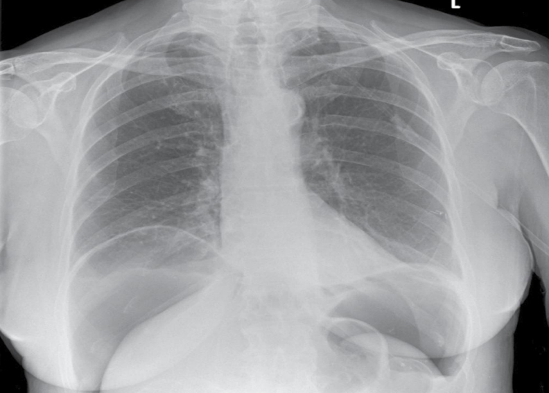
CHEST X-RAYS - CASE 2

Imaging Findings:
Chest X-ray shows a large left-sided pleural effusion with rightward mediastinal shift.
Clinical Interpretation:
Differentials include malignancy (primary/metastatic), parapneumonic effusion, TB, or post-surgical collection. Given history, malignancy is a leading concern.
Next Steps:
•Clarify prior thoracic surgery history.
•Routine blood tests.
•Start oxygen.
•Insert chest drain; send pleural fluid for cytology, biochemistry, and microbiology.
•Post-drainage chest X-ray.
•Contrast-enhanced CT thorax based on fluid analysis.
Key Learning Point:
•Massive effusions with mediastinal shift warrant urgent drainage and evaluation for malignancy
CHEST X-RAYS - CASE 3
A 65-year-old female is in the high dependency unit. She suffered a subarachnoid hemorrhage two days ago. She has
since had the culprit aneurysm coiled. You are reviewing her as she has become dyspneic and hypoxic. She has an
increased respiratory rate. Chest examination is largely unremarkable. You request a portable chest X-ray for further
assessment.
Findings
This is a portable AP VIEW erect chest X-ray in an adult.
● The patient is slightly rotated.
● There is a dense left-sided retrocardiac opacity with a linear edge. This results in the
appearance of a double left heart border. The medial aspect of the left
hemidiaphragm is not visible, indicating the pathology is within the left lower lobe.
These findings are in keeping with a left lower lobe collapse.
● There is blunting of the left costophrenic angle, suggestive of a small pleural effusion.
Imaging Findings:
Chest X-ray shows left lower lobe collapse with a small left-sided pleural effusion.
Clinical Interpretation:
The collapse is likely secondary to mucus plugging. Comparison with previous imaging is recommended
to confirm a new onset.
Next Steps:
• Initiate chest physiotherapy and supplemental oxygen.
• Review prior chest X-rays.
• Schedule follow-up chest X-ray to monitor resolution.
Key Learning Point:
Mucus plugging is a common reversible cause of lobar collapse. Early physiotherapy can help reexpand the lung
To foster collaboration between radiology and other medical specialties, ensuring a holistic approach to patient diagnosis and management.
Radiology club members collaborate with departments such as surgery, orthopedics, internal medicine, and oncology to discuss complex clinical cases. Imaging findings are correlated with clinical presentations, laboratory results, and histopathological reports. These meetings enhance interdisciplinary communication, refine diagnostic reasoning, and promote integrated patient care. By engaging in discussions with specialists from various fields, radiology residents develop a comprehensive understanding of how imaging findings influence patient management decisions, ultimately contributing to better healthcare outcomes.