Date: Dates to be communicated to members as per schedule
Aim:To provide hands-on training in basic ultrasound techniques, enabling radiology residents and medical students to develop proficiency in real-time imaging interpretation and procedural skills.
Description:
- Ultrasound is a vital diagnostic and interventional tool in modern medical practice. The “Ultrasound Live Demo & Practice” sessions are designed to provide practical exposure to real-time ultrasound imaging through direct patient interaction and simulation models.
- These sessions begin with a theoretical introduction covering ultrasound physics, image acquisition techniques, probe handling, and common artifacts. Faculty members and experienced radiologists provide demonstrations of basic ultrasound applications, including abdominal, musculoskeletal, vascular, and obstetric ultrasound.
- Participants are then divided into small groups and rotate through different ultrasound stations, where they practice hands-on scanning under expert supervision. Real patients and high-fidelity simulation models are used to mimic various clinical scenarios. Key skills taught in these sessions include:
- Identification of normal anatomical structures and tissue differentiation.
- Systematic scanning techniques for different organ systems.
- Recognition of pathological findings such as cysts, tumors, vascular abnormalities, and organ dysfunction.
- Doppler imaging techniques for assessing vascular flow and hemodynamics.
- Ultrasound-guided procedures such as central venous catheterization, biopsy guidance, and fluid drainage.
- Regular practice and expert feedback enhance trainees’ ability to obtain high-quality images, interpret findings accurately, and integrate ultrasound into clinical decision-making. This activity bridges the gap between theoretical knowledge and practical application, equipping residents with essential sonographic skills that are critical for their future practice in diagnostic and interventional radiology.
Dates to be communicated to members as per schedule
Introduction
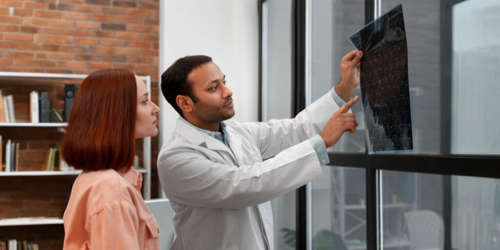
In the rapidly evolving field of radiology, proficiency in interpreting CT (Computed Tomography) and MRI (Magnetic Resonance Imaging) images is paramount for healthcare professionals. The ability to accurately analyze these imaging modalities enables clinicians to diagnose conditions effectively and formulate appropriate treatment plans. This workshop, organized by the RADNOVA radiology club, aims to enhance participants’ skills in interpreting CT and MRI images using DICOM (Digital Imaging and Communications in Medicine) viewers.
This comprehensive session will delve into the intricacies of image interpretation, focusing on the principles underlying CT and MRI technologies, the practical aspects of utilizing DICOM viewers, and the critical competencies required for effective image analysis.
Workshop Objectives
The primary objectives of this workshop are as follows:
- Understanding Imaging Modalities: Participants will gain a solid foundation in the principles of CT and MRI imaging, including their respective advantages, limitations, and clinical applications.
- Familiarization with DICOM Standards: Attendees will be introduced to the DICOM standard, which facilitates the sharing and management of medical imaging data, ensuring interoperability among different imaging devices and software.
- Hands-On Experience with DICOM Viewers: Participants will engage in hands-on sessions using DICOM viewers to explore various imaging studies, honing their skills in image manipulation and interpretation.
- Developing Analytical Skills: Through guided exercises and case discussions, attendees will learn to recognize common pathological findings, differentiate between normal and abnormal images, and enhance their overall interpretive skills.
- Integration of Clinical Context: The workshop will emphasize the importance of integrating clinical information with imaging findings to arrive at accurate diagnoses.
Workshop Outline
- Understanding Imaging Modalities
CT and MRI are essential imaging techniques that play a crucial role in modern diagnostic medicine.
CT Imaging:
Principles: CT imaging utilizes X-rays to generate cross-sectional images of the body. The X-ray tube rotates around the patient, capturing multiple images from different angles, which are then reconstructed to produce detailed images of internal structures.
Advantages: CT is particularly effective in visualizing bony structures, detecting acute hemorrhages, and assessing complex anatomical regions such as the abdomen and pelvis.
Limitations: The use of ionizing radiation poses a risk, necessitating a careful evaluation of the risks versus benefits. Additionally, CT is less effective in differentiating soft tissue contrast compared to MRI.
MRI Imaging:
Principles: MRI employs strong magnetic fields and radiofrequency pulses to generate images based on the magnetic properties of hydrogen atoms in the body. This technique produces high-resolution images of soft tissues.
Advantages: MRI excels in visualizing soft tissue structures, making it invaluable for assessing neurological, musculoskeletal, and oncological conditions.
Limitations: MRI is time-consuming, more expensive than CT, and may not be suitable for patients with certain implants or devices.
- Familiarization with DICOM Standards
The DICOM standard is essential for managing and sharing medical imaging data. Participants will learn:
DICOM Structure: Understanding the DICOM file structure, which includes metadata (patient information, imaging modality, acquisition parameters) and image pixel data.
Interoperability: The importance of DICOM in ensuring compatibility among various imaging devices and software systems, facilitating seamless data exchange.
DICOM Viewers: An overview of various DICOM viewers available for image analysis, including open-source options, commercial software, and web-based platforms.
- Hands-On Experience with DICOM Viewers
The hands-on segment of the workshop will involve interactive sessions where participants will use DICOM viewers to analyze a range of CT and MRI studies.
Image Manipulation Techniques:
Windowing and Leveling: Adjusting brightness and contrast to optimize the visualization of specific tissues.
Image Reconstruction: Understanding the reconstruction process, including 3D reconstructions and MPR (multiplanar reconstructions).
Annotation Tools: Utilizing tools for measuring distances, angles, and regions of interest (ROIs) on images.
Case Studies: Participants will be provided with a selection of clinical cases representing common and challenging scenarios in CT and MRI interpretation. They will work in groups to analyze the images, identify key findings, and discuss differential diagnoses.
- Developing Analytical Skills
Effective image interpretation requires a systematic approach. The workshop will cover:
Recognizing Normal Anatomy: Establishing a baseline understanding of normal anatomical structures as a foundation for identifying abnormalities.
Identifying Common Pathologies: Training participants to recognize prevalent conditions, such as tumors, fractures, and degenerative changes, by analyzing case examples.
Critical Thinking: Encouraging participants to develop their analytical skills through guided discussions and peer interactions, fostering a collaborative learning environment.
- Integration of Clinical Context
The final segment of the workshop will focus on integrating imaging findings with clinical context:
Clinical Correlation: Emphasizing the importance of correlating imaging results with clinical symptoms, laboratory findings, and patient history to arrive at accurate diagnoses.
Discussion of Real-World Cases: Engaging participants in discussions of real-world clinical cases where imaging played a pivotal role in diagnosis and management, highlighting the necessity of collaboration between radiologists and referring physicians.
- Conclusion
The CT & MRI Image Interpretation workshop, hosted by the RADNOVA radiology club, is designed to equip participants with essential skills and knowledge in image interpretation. By combining theoretical knowledge with practical experience using DICOM viewers, attendees will leave the workshop with enhanced confidence in their ability to analyze CT and MRI images effectively. This workshop not only aims to improve technical competencies but also fosters a deeper understanding of the vital role radiology plays in patient care.
Through this session, we hope to inspire the next generation of radiologists and healthcare professionals to continue pursuing excellence in the field of medical imaging. As the landscape of radiology evolves, ongoing education and hands-on experience will remain critical in ensuring that practitioners are well-prepared to meet the challenges of modern medicine.
- References
American College of Radiology (ACR). (2023). ACR Appropriateness Criteria.
DICOM Standards Committee. (2023). DICOM Standard.
Brant, W. E., & Helms, C. A. (2012). Fundamentals of Diagnostic Radiology (4th ed.). Lippincott Williams & Wilkins.
Latchaw, R. E., et al. (2009). “American College of Radiology White Paper on Imaging and Informatics.” Journal of the American College of Radiology
Dates to be communicated to members as per schedule
Introduction
Interventional Radiology (IR) is a subspecialty of radiology that utilizes minimally invasive procedures to diagnose and treat a variety of medical conditions. By employing imaging guidance techniques, such as fluoroscopy, ultrasound, CT, and MRI, interventional radiologists can perform a wide range of procedures with precision and reduced patient risk. This workshop, organized by the RADNOVA radiology club, aims to provide participants with a comprehensive introduction to basic interventional radiology procedures, such as biopsies and catheter placements.
The workshop will cover the fundamental principles of interventional radiology, explore various IR techniques and their clinical applications, and provide hands-on training in basic procedures. By the end of the session, participants will be equipped with essential knowledge and skills to appreciate the role of IR in modern medicine and enhance their competency in performing basic IR procedures.
Workshop Objectives
The primary objectives of this workshop are as follows:
- Understanding Interventional Radiology: Participants will gain an understanding of the principles and practices of interventional radiology, including the various imaging modalities used in IR.
- Familiarization with Basic Procedures: Attendees will learn about common interventional radiology procedures, such as biopsies, catheter placements, and drainage procedures, including their indications and contraindications.
- Hands-On Experience: Participants will engage in hands-on training sessions to practice basic IR techniques using simulators or models, enhancing their procedural skills.
- Exploration of Clinical Applications: The workshop will emphasize the clinical applications of IR procedures in various medical fields, including oncology, vascular disease, and gastroenterology.
- Patient Safety and Ethical Considerations: Participants will learn about the importance of patient safety, informed consent, and ethical considerations in interventional radiology practice.
Workshop Outline
- Understanding Interventional Radiology
Interventional radiology has transformed the landscape of modern medicine by providing minimally invasive alternatives to traditional surgical procedures. Participants will be introduced to the following topics:
Definition and History of Interventional Radiology:
Overview of the evolution of interventional radiology as a subspecialty, highlighting key milestones in its development and the introduction of various imaging technologies.
Principles of Image Guidance:
Understanding the role of imaging modalities in guiding interventional procedures, including:
Fluoroscopy: Real-time X-ray imaging for visualizing dynamic processes, particularly in vascular interventions.
Ultrasound: Utilizing sound waves to visualize soft tissue structures and guide needle placement in biopsies and drainage procedures.
CT and MRI: Providing high-resolution imaging for accurate localization of lesions and guiding complex interventions.
The Role of Interventional Radiologists:
Exploring the multifaceted roles of interventional radiologists in healthcare, including diagnosis, treatment, and collaboration with other specialties.
- Familiarization with Basic Procedures
This section will focus on the most commonly performed interventional radiology procedures, their indications, contraindications, and techniques. Participants will learn about:
Biopsy Procedures:
Indications: Understanding when biopsies are necessary for diagnosing malignancies or infections.
Techniques: Overview of various biopsy techniques, including fine-needle aspiration (FNA), core needle biopsy, and image-guided biopsies.
Procedural Steps:
Patient preparation, including obtaining informed consent and ensuring appropriate imaging guidance.
Techniques for local anesthesia and sedation, as well as the use of ultrasound or CT for needle localization.
Catheter Placement Procedures:
Types of Catheters: Understanding various catheter types, including central venous catheters (CVCs), peripherally inserted central catheters (PICCs), and drainage catheters.
Indications: Situations where catheter placement is indicated, such as for chemotherapy, long-term medication administration, or drainage of abscesses.
Procedural Steps:
Selection of appropriate access site and imaging guidance for catheter placement.
Techniques for catheter insertion, securing the catheter, and post-procedure care.
Drainage Procedures:
Indications: Understanding when percutaneous drainage is necessary for abscesses, fluid collections, or biliary obstruction.
Techniques: Overview of drainage techniques and the importance of imaging guidance to ensure accurate placement.
- Hands-On Experience
The hands-on portion of the workshop is designed to provide participants with practical experience in basic interventional radiology procedures. Participants will engage in simulated training sessions where they will:
Practice Biopsy Techniques:
Utilizing models or simulators to practice performing biopsies, including needle insertion, localization, and obtaining tissue samples.
Learn Catheter Placement Skills:
Engaging in hands-on training for catheter placement using simulation models, focusing on proper technique, securing the catheter, and troubleshooting common issues.
Explore Drainage Procedures:
Simulating drainage procedures to gain proficiency in identifying fluid collections and placing drainage catheters.
Feedback and Assessment:
Participants will receive feedback on their techniques from experienced instructors, enhancing their skills and confidence in performing these procedures.
- Exploration of Clinical Applications
This section of the workshop will focus on the various clinical applications of interventional radiology procedures in different medical fields:
Oncology:
The role of interventional radiology in cancer diagnosis and treatment, including tumor biopsies, radiofrequency ablation, and transarterial chemoembolization (TACE).
Vascular Disease:
Understanding how IR procedures are used to treat vascular conditions, such as peripheral artery disease, thrombolysis for deep vein thrombosis, and stenting.
Gastroenterology:
Exploring the use of interventional radiology in managing gastrointestinal conditions, including biliary drainage, portosystemic shunts, and feeding tube placements.
Urology:
The applications of IR in urological conditions, such as nephrostomy tube placement and renal biopsy.
Emergencies:
Discussing the critical role of interventional radiology in emergency settings, including trauma management and acute gastrointestinal bleeding interventions.
- Patient Safety and Ethical Considerations
Patient safety is paramount in interventional radiology. This segment will address:
Risk Assessment and Management:
Understanding potential complications associated with interventional procedures and strategies for minimizing risks.
Informed Consent:
The importance of obtaining informed consent, ensuring that patients understand the procedure, its risks, and benefits.
Ethical Considerations:
Discussing the ethical responsibilities of interventional radiologists, including patient autonomy, beneficence, and non-maleficence.
Quality Improvement and Best Practices:
Emphasizing the need for continuous quality improvement in interventional radiology, including adherence to guidelines and protocols to enhance patient safety.
Conclusion
The Basic Interventional Radiology workshop organized by the RADNOVA radiology club serves as an essential platform for medical students, residents, and healthcare professionals to gain a foundational understanding of interventional radiology procedures. By integrating theoretical knowledge with hands-on experience, participants will enhance their skills and confidence in performing basic IR techniques, such as biopsies and catheter placements.
This workshop highlights the significance of interventional radiology as a critical component of modern medicine, illustrating its diverse applications across various clinical specialties. As the field of interventional radiology continues to evolve, ongoing education and training will remain vital in ensuring practitioners are equipped to provide safe and effective care to their patients.
References
American College of Radiology (ACR). (2023). ACR Appropriateness Criteria.
Sacks, D., et al. (2010). “Interventional Radiology: A Guide to the Specialty.” American Journal of Roentgenology.
Dake, M. D., et al. (2017). “Interventional Radiology: Clinical Applications.” In Interventional Radiology: A Practical Guide (pp. 1-15). Springer.
Becker, G. J., & Kuhlman, J. E. (2016). “Safety and Effectiveness of Interventional Procedures.” Radiology.
Nascimento, F. A., et al. (2018). “The Role of Interventional Radiology in Emergency Medicine.” Emergency Medicine Clinics of North America.
Introduction
Fluoroscopy plays a vital role in modern diagnostic radiology by providing real-time, dynamic imaging of internal structures. Among its many applications, gastrointestinal (GI) contrast studies using barium remain a cornerstone for evaluating structural and functional abnormalities of the digestive system. These studies allow radiologists and clinicians to assess swallowing mechanisms, esophageal motility, gastric emptying, small bowel transit, and colonic function in both routine and complex clinical scenarios.
This workshop, organized by the RADNOVA radiology club, is designed to provide an in-depth understanding of fluoroscopic techniques, barium-based contrast studies, and their interpretation. It aims to equip participants with the necessary knowledge and practical skills to accurately assess GI pathology using contrast fluoroscopy.
Workshop Objectives
The primary objectives of this workshop include:
- Understanding Fluoroscopy Principles – Exploring the fundamental physics, equipment, and safety considerations in fluoroscopic imaging.
- Learning Barium-Based Contrast Studies – Understanding indications, techniques, and protocols for conducting barium swallow, upper GI series, small bowel follow-through (SBFT), and barium enema studies.
- Image Interpretation Skills – Training in systematic evaluation of fluoroscopic images and recognizing normal and abnormal findings.
- Dynamic Imaging Techniques – Learning how real-time imaging enhances the diagnosis of motility disorders and functional abnormalities.
- Clinical Correlation – Integrating fluoroscopic findings with clinical presentations to optimize patient management.
- Radiation Safety & Optimization – Understanding dose reduction strategies and the importance of ALARA (As Low As Reasonably Achievable) principles in fluoroscopy.
By the end of the workshop, participants will have a strong foundation in dynamic imaging of the GI tract and will be able to systematically approach fluoroscopic studies in clinical practice.
Workshop Outline
- Fundamentals of Fluoroscopy
Fluoroscopy provides continuous X-ray imaging in real-time, enabling the visualization of dynamic processes such as swallowing, gastric emptying, and bowel peristalsis. Participants will be introduced to:
Principles of Fluoroscopy – Understanding how X-ray beams interact with contrast agents to create moving images.
Fluoroscopic Equipment & Technology – Exploring image intensifiers, digital fluoroscopy, pulsed fluoroscopy, and radiation exposure considerations.
Indications & Contraindications – Identifying when fluoroscopy is the preferred imaging modality and recognizing contraindications such as suspected bowel perforation or aspiration risk.
Radiation Dose Considerations – Learning about exposure minimization strategies, including collimation, dose optimization, and protective shielding.
- Overview of GI Contrast Agents
Barium-based contrast agents are widely used in fluoroscopic GI studies due to their excellent mucosal coating and high radiopacity. The session will cover:
Types of Contrast Agents:
Barium Sulfate – The standard agent for GI fluoroscopy due to its non-absorbable nature.
Water-Soluble Iodinated Contrast – Used in cases of suspected perforation or aspiration risk.
Thick vs. Thin Barium – Choosing the appropriate contrast consistency based on clinical indications.
Mechanisms of Contrast Transit: Understanding how different viscosities of barium affect swallowing mechanics, gastric emptying, and intestinal transit.
Complications & Safety: Discussion on rare but serious complications such as aspiration pneumonia, contrast-induced perforation, and barium impaction.
- Barium Swallow &Esophagography
The barium swallow is a fundamental study used to evaluate the esophagus. This section will include:
Indications:
Dysphagia (difficulty swallowing)
Suspected strictures or masses
Evaluation of gastroesophageal reflux disease (GERD)
Assessment of motility disorders (e.g., achalasia, diffuse esophageal spasm)
Technique:
Proper patient positioning (upright, prone, supine)
Use of single vs. double contrast techniques
Real-time assessment of bolus transit and peristalsis
Interpretation:
Normal esophageal anatomy and motility
Abnormal findings:
Strictures, webs, and rings (e.g., Schatzki’s ring)
Achalasia – “Bird-beak” appearance
Zenker’s diverticulum
Esophageal varices – serpiginous filling defects
Carcinoma – irregular, shouldered narrowing
- Upper Gastrointestinal (UGI) Series
The upper GI series evaluates the stomach and duodenum, often complementing endoscopic findings. Topics covered will include:
Indications:
Peptic ulcer disease
Gastric outlet obstruction
Hiatal hernia
Malrotation or volvulus
Technique:
Administration of effervescent granules for double-contrast imaging
Prone, supine, oblique positioning for optimal gastric visualization
Real-time fluoroscopy to assess motility
Interpretation:
Normal anatomy and gastric emptying patterns
Pathological findings:
Peptic ulcers – focal outpouching with radiating folds
Gastric malignancies – irregular, non-distensible mass with ulceration
Hiatal hernia – gastric fundus above the diaphragm
Pyloric stenosis – “String sign” with delayed emptying
- Small Bowel Follow-Through (SBFT) &Enteroclysis
Small bowel imaging is crucial for assessing malabsorption, Crohn’s disease, and mechanical obstruction. This segment will cover:
Indications:
Unexplained diarrhea or malabsorption
Suspected Crohn’s disease
Small bowel obstruction (SBO) evaluation
Technique:
Serial imaging at 15-30 min intervals post-barium ingestion
Trendelenburg positioning for enhanced visualization of jejunal loops
Interpretation:
Normal peristalsis and fold pattern
Crohn’s disease – “String sign” due to luminal narrowing
Small bowel obstruction – dilated loops with air-fluid levels
Celiac disease – mucosal flattening, segmentation
- Barium Enema (Lower GI Studies)
The barium enema is instrumental in evaluating colonic pathology. This session will include:
Indications:
Colonic polyps and neoplasms
Diverticular disease
Chronic constipation or obstruction
Technique:
Single-contrast vs. double-contrast methods
Patient positioning for optimal opacification
Air insufflation techniques for enhanced mucosal visualization
Interpretation:
Diverticulosis – outpouchings along colonic wall
Polyps and masses – sessile or pedunculated filling defects
Apple-core lesion – classic appearance of colonic malignancy
Volvulus – “Bird’s beak” tapering at the twisted bowel segment
- Dynamic Imaging & Functional Assessment
One of the key advantages of fluoroscopy is its ability to assess real-time function. Thissection will emphasize:
Swallowing disorders – Videofluoroscopic swallowing studies (VFSS)
Gastroesophageal reflux – Real-time detection of acid reflux episodes
Colonic transit studies – Evaluating slow-transit constipation or Hirschsprung’s disease
Conclusion
This workshop offers a comprehensive introduction to fluoroscopic GI contrast studies,equipping participants with essential skills in image acquisition, interpretation, and clinical decision-making. By integrating hands-on training with case-based discussions, the RADNOVA radiology club aims to foster a deeper appreciation for fluoroscopic imaging and its crucial role in patient care.
Whether pursuing a career in radiology, gastroenterology, or general medicine, attendees will gain valuable insights into the dynamic world of GI imaging, enhancing their ability to diagnose and manage a wide spectrum of digestive disorders.
References
Levine, M. S., & Laufer, I. (2011). Gastrointestinal Radiology: A Pattern Approach.
Maglinte, D. D. T., et al. (2015). Radiologic Evaluation of the Small Bowel Disorders.
American College of Radiology (ACR) Guidelines for Gastrointestinal Fluoroscopy
Introduction
Portable imaging plays a crucial role in the management of critically ill patients in the Intensive Care Unit (ICU) and Emergency Room (ER). In settings where patient mobility is limited, bedside radiography and ultrasound (USG) serve as invaluable tools for rapid diagnosis and intervention. Portable X-ray allows clinicians to assess life-threatening conditions such as pneumothorax, pulmonary edema, fractures, and central line placements, while bedside ultrasound provides real-time dynamic imaging for hemodynamic assessment, fluid status evaluation, and procedural guidance.
This workshop, organized by the RADNOVA radiology club, is designed to provide hands-on experience in interpreting portable radiographs and performing point-of-care ultrasound (POCUS) in critically ill patients. Participants will learn about the technical aspects, common pathologies encountered in the ICU/ER, and the integration of these imaging modalities into clinical decision-making.
Workshop Objectives
By the end of this workshop, participants will be able to:
- Understand the Principles of Portable Imaging – Learn about the physics, equipment, and technical limitations of portable X-ray and bedside ultrasound.
- Interpret Portable Chest & Abdominal Radiographs – Develop a systematic approach to analyzing portable X-rays, recognizing normal and pathological findings in ICU and ER settings.
- Perform Bedside Ultrasound (POCUS) Techniques – Gain hands-on experience in focused ultrasound applications, including lung, cardiac, abdominal, and vascular assessment.
- Identify Critical ICU & ER Pathologies – Recognize life-threatening conditions such as pneumothorax, pulmonary edema, free intraperitoneal fluid, and cardiac tamponade.
- Enhance Procedural Skills with USG Guidance – Learn how to use ultrasound for vascular access, thoracentesis, paracentesis, and other bedside procedures.
- Understand Clinical Integration & Decision-Making – Apply imaging findings to optimize patient management in emergency and critical care settings.
This workshop aims to bridge the gap between imaging and clinical medicine by empowering participants with the knowledge and skills to interpret and perform bedside imaging effectively.
Workshop Outline
Principles of Portable Imaging in Critical Care
Portable imaging differs from standard radiographic studies due to several technical and environmental challenges. This section will cover:
Physics and Technical Considerations
Mobile X-ray equipment and radiation safety precautions
Limitations of portable imaging (e.g., patient positioning constraints, motion artifacts)
Image quality optimization strategies
Indications for Portable X-ray & Ultrasound in ICU/ER
Assessing respiratory distress, cardiovascular compromise, and acute abdominal conditions
Monitoring disease progression and response to treatment
Procedural guidance (e.g., central line placement, pleural drainage)
- Portable Chest X-ray (CXR) Interpretation
Portable chest radiographs are among the most commonly performed imaging studies in critically ill patients. This session will focus on:
Systematic Approach to CXR Interpretation (ABCDE Method)
Airway – Tracheal position, endotracheal tube (ETT) placement
Breathing – Lung expansion, consolidation, pneumothorax
Circulation – Cardiomegaly, pulmonary edema
Diaphragm – Free air (pneumoperitoneum), diaphragmatic elevation
Everything Else – Lines, tubes, fractures, foreign bodies
Common ICU & ER Findings on Portable CXR
Endotracheal Tube (ETT) Placement – Assessing proper positioning at T2-T4
Nasogastric (NG) Tube Placement – Verifying tube position and detecting malposition
Central Venous Catheter (CVC) & PICC Line Positioning – Avoiding complications such as arterial puncture or pneumothorax
Pulmonary Edema – “Bat-wing” pattern, Kerley B lines
Pneumothorax – Deep sulcus sign, absence of lung markings
Pleural Effusion – Meniscus sign, subpulmonic effusion
ARDS (Acute Respiratory Distress Syndrome) – Bilateral opacities, ground-glass changes
COVID-19 & Viral Pneumonia Findings – Peripheral, bilateral ground-glass opacities
- Portable Abdominal X-ray Interpretation
Portable abdominal X-rays are crucial in detecting acute gastrointestinal and surgical emergencies. This session will include:
Indications for Portable Abdominal X-ray
Suspected bowel perforation (pneumoperitoneum)
Bowel obstruction and ileus
Foreign body ingestion
Ascites and organomegaly assessment
Systematic Approach to Interpretation
Gas Patterns – Normal vs. abnormal gas distribution
Pneumoperitoneum – Free air under the diaphragm, Rigler’s sign
Bowel Obstruction – Dilated loops, air-fluid levels, transition point
Ileus vs. Mechanical Obstruction – Diffuse gas distribution vs. localized dilatation
Calcifications – Gallstones, renal stones, pancreatic calcifications
- Bedside Ultrasound (POCUS) Training
Point-of-care ultrasound (POCUS) has revolutionized bedside diagnosis and procedural guidance. This hands-on session will cover:
- Basic Ultrasound Physics & Knobology
Understanding probe selection:
Linear probe – Vascular access, soft tissue
Curvilinear probe – Abdominal, FAST exam
Phased-array probe – Cardiac, lung ultrasound
Machine settings, gain adjustment, depth selection, and image orientation
- Lung Ultrasound (LUS) in the ICU
Lung ultrasound is more sensitive than chest X-ray for detecting certain conditions. Participants will learn:
Normal Lung Patterns – A-lines, lung sliding
Pathological Findings
Pneumothorax – Absent lung sliding, barcode sign
Pleural Effusion – Anechoic collection, spine sign
Pulmonary Edema – B-lines (comet-tail artifacts)
- Cardiac Ultrasound (FOCUS/Echo in Shock)
Bedside echocardiography is essential for assessing cardiac function in unstable patients. This session includes:
Basic Cardiac Views
Parasternal long axis, apical four-chamber, subxiphoid view
Rapid Cardiac Assessment
Pericardial effusion & tamponade (right ventricular collapse)
Left ventricular function (global hypokinesia in shock)
Inferior vena cava (IVC) collapsibility for fluid responsiveness
- Abdominal Ultrasound & FAST Exam
Focused Assessment with Sonography for Trauma (FAST) is a rapid bedside scan for internal bleeding. Key components:
RUQ (Morrison’s Pouch) – Liver-kidney interface
LUQ (Splenorenal View) – Spleen-kidney interface
Pelvic View (Pouch of Douglas) – Free fluid detection
Pericardial View – Tamponade assessment
- Vascular & Procedural Ultrasound
Ultrasound-Guided Vascular Access
Central line insertion (IJ, subclavian, femoral)
Peripheral IV access in difficult cases
Procedural Guidance
Thoracentesis, paracentesis
Joint aspiration, nerve blocks
- Clinical Integration & Case-Based Discussion
Real-life ICU and ER cases will be presented, allowing participants to:
Correlate Imaging Findings with Clinical Presentations
Discuss Management Strategies Based on Imaging Results
Practice Decision-Making in Acute Settings
Conclusion
This workshop provides an immersive learning experience in portable X-ray interpretation and bedside ultrasound, emphasizing their critical role in ICU and ER settings. By combining theoretical knowledge with hands-on practice, participants will develop confidence in interpreting portable imaging studies and performing bedside ultrasound, ultimately improving patient care in emergency and critical care environments.
Whether you are a medical student, resident, or practicing physician, mastering these essential imaging skills will enhance your ability to make rapid, informed decisions in high-stakes clinical situations.
References
Lichtenstein, D. A. (2014). Lung Ultrasound in the Critically Ill.
American College of Radiology (ACR) Guidelines on Portable Imaging.
Blaivas, M. (2020). Point-of-Care Ultrasound in Critical Care Medicine.